
Teratoma quístico maduro abdominal: Hallazgos imagenológicos y espécimen quirúrgico
Abdominal mature cystic teratoma: Imaging findings and surgical specimen
DOI:
https://doi.org/10.17268/rmt.2023.v18i3.5171Resumo
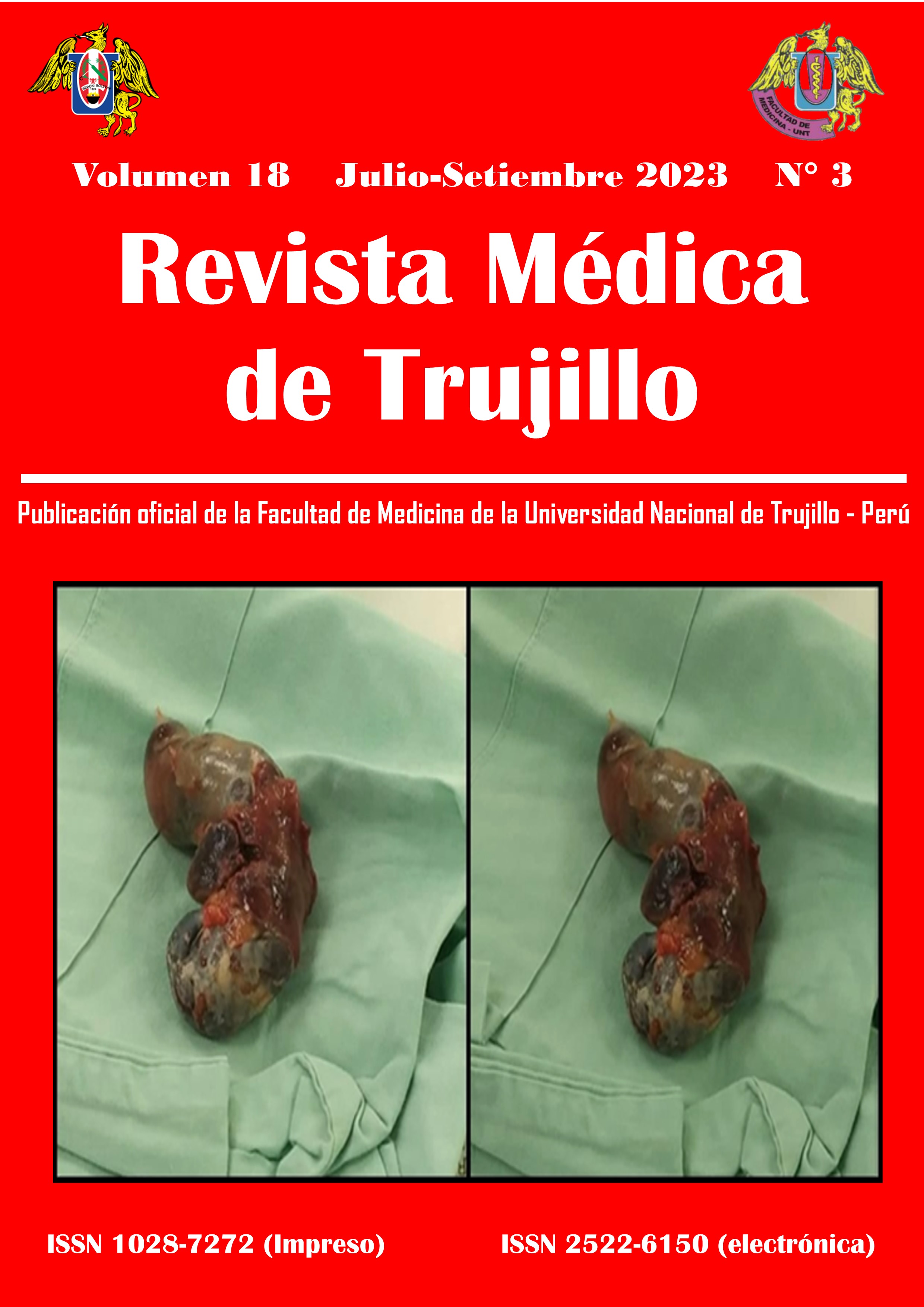
Paciente femenina de 3 meses con antecedente de hernia inguinal que ingresa a hospitalización por síndrome febril. Al examen físico con distensión abdominal, en su estancia hospitalaria se realizó RNM de abdomen y pelvis contrastada con gadolinio (A) con hallazgos masa intraperitoneal en el flanco izquierdo, mesogastrio y epigastrio, de aspecto quístico, de paredes delgadas, con componente sólido en su interior, con pequeñas zonas hiperintensas que presentan supresión de la señal en las secuencias con saturación grasa, que puede corresponder a componente lipomatoso, con efecto de masa, asociando desplazamiento de las asas intestinales delgadas y del colon, sin infiltración de órganos adyacentes, a considerar la posibilidad de teratoma quístico maduro, con dimensiones aproximadas de 10,9 x 8,7 x 10,9 cm (L x AP x T). Posteriormente se lleva a resección por departamento de cirugía pediátrica, muestra histopatológica de espécimen quirúrgico(B) compatible con teratoma quístico maduro.
Publicado
Como Citar
Edição
Seção
Licença
Copyright (c) 2023 Revista Médica de Trujillo

Este trabalho está licenciado sob uma licença Creative Commons Attribution-NonCommercial 4.0 International License.