Inhibitory effect of valeryl chloride-acylated blueberry (Vaccinium corymbosum) anthocyanins on lipid oxidation in UHT milk
Efecto inhibidor de las antocianinas de arándano (Vaccinium corymbosum) aciladas con cloruro de valerilo sobre la oxidación lipídica en leche UHT
Angie Carbajal Rivera1; Olivio Nino Castro Mandujano1
2 Universidad Nacional Mayor de San Marcos, Lima, Peru.
ORCID de los autores:
A. Carbajal Rivera: https://orcid.org/0009-0005-1287-1676 O. N. Castro Mandujano: https://orcid.org/0000-0002-6592-6934
ABSTRACT
Food oxidation is a prevalent issue that deteriorates nutritional and organoleptic values, potentially compromising consumer health. A specific concern is the oxidation of fatty acids in UHT cow’s milk. This research evaluates the fatty acid oxidation inhibition capacity of acylated and non-acylated anthocyanin extracts derived from blueberries (Vaccinium corymbosum). The anthocyanins were obtained through alcoholic extraction and subsequently acylated using valeryl chloride. The extracts were characterized via FTIR-ATR spectroscopy, UV-Vis spectrophotometry, and antioxidant capacity using the DPPH assay. Additionally, the acylation reaction mechanism was determined through computational modeling in Gaussian. Results showed a higher degree of inhibition in the acylated anthocyanin extract. This was further confirmed through organoleptic testing of UHT milk samples and the evaluation of the fatty acid profile via GC-FID.
Keywords: Acylated anthocyanins; Antioxidant; UHT milk; Lipid oxidation; Computational chemistry.
RESUMEN
La oxidación de los alimentos es un problema prevalente que deteriora los valores nutricionales y organolépticos, comprometiendo potencialmente la salud del consumidor. Una preocupación específica es la oxidación de los ácidos grasos en la leche de vaca UHT. Esta investigación evalúa la capacidad de inhibición de la oxidación de ácidos grasos de extractos de antocianinas aciladas y no aciladas derivadas de arándanos (Vaccinium corymbosum). Las antocianinas se obtuvieron mediante extracción alcohólica y posteriormente se acilaron utilizando cloruro de valerilo. Los extractos se caracterizaron mediante espectroscopía FTIR-ATR, espectrofotometría UV-Vis y capacidad antioxidante mediante el ensayo DPPH. Adicionalmente, el mecanismo de la reacción de acilación se determinó a través de modelado computacional en Gaussian. Los resultados mostraron un mayor grado de inhibición en el extracto de antocianina acilada. Esto se confirmó posteriormente mediante pruebas organolépticas de muestras de leche UHT y la evaluación del perfil de ácidos grasos a través de GC-FID.
Palabras clave: Antocianinas aciladas; Antioxidante; leche UHT; Oxidación lipídica; Química computacional.
1. Introduction
The application of acylated anthocyanins has been studied as a strategy to inhibit fatty acid lipid oxidation and thus extend the shelf life of UHT milk through the use of a natural preservative. Anthocyanins are phenolic compounds widely recognized for their antioxidant capacity, which increases the significant potential for food applications. To modify their solubility, an acylation of anthocyanins extracted from the Vaccinium corymbosum fruit was performed as the study of Wang et al. (2022) and Teng et al. (2022). The purpose of this application is to mitigate the oxidation process of lipids present in UHT (Ultra-High Temperature) cow's milk, as oxidized lipids can cause health issues, and the excessive use of synthetic additives may also be detrimental to human health. Blueberries are an excellent source of phenolic compounds; these phytochemicals are widely distributed across plant-based materials, possess known antioxidant capabilities, and are present in high concentrations in blueberries according to Vyas et al. (2013). Anthocyanin extracts were extracted and characterized from Vaccinium corymbosum, and the anthocyanin extract was acylated and characterized through a reaction with valeryl chloride. Subsequently, the ability of these modified compounds to inhibit the oxidation of fatty acids in UHT milk was evaluated, aiming to explore and implement them as a natural additive to utilize their inhibitory effect on fatty acid oxidation in dairy products or emulsions.
2. Methodology
2.1 Extraction and characterization of antho-cyanins from the Vaccinium corymbosum
For the extraction of anthocyanins, 50 g of blueberries from Ancash, Peru were used and were ground and mixed with 150 g of a solvent of ethanol acidified with 1% citric acid (1:3 sample/solvent), then subjected to ultrasound at 36 ºC, and afterwards filtered and the liquid extract was separated from the solid. Then the extract was concentrated at 40 ºC because it is heat-sensitive and it has been reported that using higher temperatures leads to a loss of the concentration of anthocyanins present; if heated to 60 ºC, half of the anthocyanin concentration is lost (Liu et al., 2018) The extract was stored at approximately 4 ºC. Finally, the extract was dried in an oven at 41 ºC for 7 days to remove traces of solvent, where a viscous mass was obtained that was subsequently used as a preservative in sample M2 (Table 4).
The content of total anthocyanins was analyzed, the determination and quantification of antho-cyanins present in the extract was carried out using the pH differential method in a UV-Vis spectrophotometer, this method is based on the structural change of anthocyanins when the pH is modified (pH 1 colored and pH 4.5 colorless). Dilutions of the alcoholic anthocyanin extract were prepared with a pH 4.5 sodium acetate buffer solution and with a pH 1.0 potassium chloride buffer solution. A measu-rement of the absorbance of all the samples is made at the wavelength of maximum absorbance (λmax=517 nm) and at 700 nm (Gulcin & Alwasel. 2023). The content of anthocyanins was expressed in mg of cyanidin-3-glucoside/100 mL.
2.2 Synthesis and characterization of acylated anthocyanins from Vaccinium corymbosum
For the synthesis 2.297 grams of solid from the previously ground blueberry extract were weighed with a small amount of ethanol as a solvent; then 1.670 grams of valeryl chloride reagent and 0.226 grams of pyridine, which acts as the catalyst, were weighed and placed in a polytetrafluoroethylene reactor at 50 ºC for approximately 24 hours. Then the mixture was washed with tetrahydrofuran-THF and filtered to remove 4-dimethylaminopyridine and the unreacted acyl chloride. The reaction product was dried at 50 ºC to remove residual tetrahydro-furan. The characterization of the acylated extract was carried out by FTIR-ATR spectroscopy (Fourier Transform Attenuated Total Reflectance Infrared).
2.3 Computational simulation of the acylation reaction
Calculations of transition states between the cya-nidin-3-o-glucoside structure and valeric acid were carried out. The calculations performed are based on proton affinity, where an isodesmic reaction was generated, a reaction in which the number of bonds and lone electron pairs are equal in reactants and products. Reaction simulations were carried out in different positions using the Gaussian platform with the functional B3lyp/ 631G(d); and the activation energy of point 1, 6, 8 was calculated where lower activation energy was observed at point 1 of the -OH (Figure 1) with 4.08 kcal/mol.
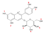
Figure 1. Cyanidin 3-glucoside structure.
2.4 Analysis of the oxidative inhibition capa-city of acylated anthocyanin extract in the fatty acids of UHT milk
To measure the antioxidant capacity of the extracts, the test with 2.2-diphenyl-1-picry-lhydrazyl (DPPH) was carried out to determine the antiradical activity. The reaction between DPPH and an antioxidant can be represented as follows:
[DPPH*] + [AOH] → [DPPH-H] + [AO*]
Concentrations of the standards between 0, 5, 10, 15, 20, 30 and 40 μL of ascorbic acid, and DPPH solution at 40 ppm dissolved in methanol were prepared for the calibration curve. Then, 4 vials with concentrations of 50, 100, 150 and 200 μL were prepared to analyze the antioxidant capacity of the extract. The reaction was carried out in the dark for 30 minutes at a temperature of 22 °C; the optical density was read on a UV-Vis spectro-photometer, where at 516 nm wavelength an increase in signal was observed (Lock, 2016)
A fat extraction from UHT milk was carried out using the AOAC 996.06 (2023) method to proceed with the analysis of free fatty acids by gas chromatography with an FID detector. A combined hydrolysis and ether extraction was performed. Finally, the concentration percentages of the different samples of fresh UHT milk (time zero), UHT milk without additives, UHT milk with anthocyanins, and UHT milk with acylated anthocyanins were compared over time.
3. Results and discussion
3.1 Characterization of anthocyanin extracts
From the extract, an absorption peak at 524 nm was obtained in the UV-Vis spectrophotometer, close to that reported by Falcone et al. (2025). A solution was prepared with the anthocyanin extract and 96º ethanol. Ethanol was used as a blank and it was scanned from 200 to 700 nm (UV-Vis spectrophotometry technique). Three peaks were obtained: a peak at 524 nm with an absor-bance of 0.340 and also peaks at 284 and 208 nm.
While the solid extract was left to dry at 43ºC for a week and was characterized by FTIR-ATR (Johnson et al., 2022), the results of the FTIR-ATR spectroscopy characterization (Table 1; Figure 2).
Table 1
FTIR-ATR results of the Solid Anthocyanin Extract
Wavelength cm-1 | Functional Groups |
3400-3200 2915-2849 1733 1623 1004 | -OH C-H and OH Double bond C=O CH and aromatics Fingerprint |
C=O absorption features were obtained between 1733 cm-1, aromatics between 1645 cm-1 and 1623 cm-1, and the -OH at 3400-3200 cm-1, which indicate the presence of these functional groups’ characteristic of the structure of an anthocyanin.
3.2 Determination of total anthocyanins
A pH 1 and pH 4.5 buffer was prepared with KCl and HCl, and sodium acetate and acetic acid, respectively. The solutions were prepared in a 2:8 ratio of sample to buffer, respectively, and readings were taken using the VISIONpro software.
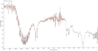
Figure 2. Solid anthocyanin extract spectrum.
The determination and quantification of antho-cyanins present in the extract was carried out using the differential pH method, which is based on the structural change of anthocyanins when the pH is modified at pH 1 it is colored and pH 4.5 colorless (Taghavi et al., 2020). The dilutions of the alcoholic anthocyanin extract were prepared with a pH 4.5 sodium acetate buffer solution and with a pH 1.0 potassium chloride buffer solution. The absorbance of all samples was measured at the wavelength of maximum absorbance (λmax = 515 nm) and at 700 nm; and a total of 178.21 mg/L of anthocyanins was obtained in the extract.
3.3 Identification reactions
Two tests were carried out in triplicate in test tubes with 1 mL of the anthocyanin extract and 1 mL of reagent. Addition of alkali to identify phenolates: NaOH 20%: A color change occurred, obtaining a dark green color. This indicates alkaline pheno-lates that are formed in the anthocyanins:
Anthocyanins–OH + NaOH →
Anthocyanins–O⁻Na⁺+ H₂O
Upon the addition of 5% lead acetate, the forma-tion of a bluish-purple precipitate was observed. This reaction indicates the formation of a lead-anthocyanin complex (lead salt), a characteristic qualitative test confirming the presence of these phenolic pigments:
Anthocyanins-(OH)n + Pb(CH₃COO)₂ → [Anthocyanins–OnPb] + 2CH₃COOH
3.4 Phytochemical analysis
A phytochemical screening was carried out based on the Lock (2016) procedure, modified. The screening was performed with the blueberry anthocyanin extract with identification reactions for the following secondary metabolites: alkaloids, flavonoids, tannins, steroids, and anthraquinones.
A reddish precipitate was obtained for the Dragendorff reaction, for the Mayer reaction (Mercury and Potassium iodide) a whitish precipitate; while the Ehrlich reaction showed a violet coloration, in the alkaloid identification reactions. For the identification of flavonoids, the Shinoda test was performed, in which a reddish coloration was obtained. In the identification of tannins, the test with FeCl3 and gelatin was performed, in which a dark green coloration for phenols and a precipitate, respectively, were obtained according to the expected results (Godlewska et al., 2023). Coloration tests were also prepared that gave negative results for steroids, anthraquinones, and saponins.
3.5 DPPH antioxidant capacity analysis
The antioxidant potential was evaluated through the DPPH radical scavenging assay. The initial absor-bance of the control was 2.960, reflecting the high reactivity of the system. The extracts demonstrated significant antioxidant capacity, with inhibition of 94.26%, 95.95%, 96.97%, and 97.77% correspon-ding to the evaluated concentrations (Table 2). These results indicate a dose-dependent response, with the highest concentration achieving the maxi-mum inhibition of 97.77% corresponding to 200 μL.
Table 2
DPPH Absorbance
Concentration μL | Absorbance |
50 100 150 200 | 0.066 0.094 0.120 0.170 |
3.6 Acylation of anthocyanin extract from blueberry
The product formed by the synthesis or acylation was characterized by FTIR-ATR spectroscopy. A decrease in the intensity of the -OH band for the hydroxyl group and an increase in the intensity of the C=O groups were observed compared to the results of the characterization of the solid anthocyanin extract by FTIR-ATR before being acylated, which could confirm the acylation process of the anthocyanins or oxidation of the -OH group, an index carbonyl was also calculated with these results. A band around 800 cm-1 for -Cl, around 300 cm-1 for -CH, and around 1700 cm-1 for the carbonyl C=O were also observed (Table & Figure 3).
Table 3
FTIR-ATR results of acylated anthocyanin extract
Wavelength cm-1 | Functional groups |
3359 2917 1736 1636 1028 | -OH C-H and OH Double bond C=O CH and aromatics Fingerprint |
The carbonyl index is used to rate the degree of oxidation of a polymer. The carbonyl index (CI) is calculated where the height or area of the carbonyl peak is normalized to the height or area of a stable reference peak (Celina et al., 2021). The intensity of reference from the group -CH was obtained at 1636 cm-1 and the intensity of the carbonyl group obtained at 1736 cm-1 gave a result of CI=1.27 with indicates the effective oxidation process, the cal-culation was made according to Yang et al. (2023).
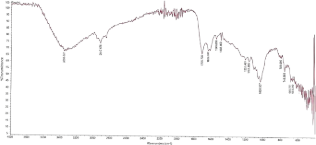
Figure 3. Acylated anthocyanin extract spectrum.
Further studies might be performed to quantify the acylation with HPLC-MS. Additionally, the extract showed a loss of solubility in water and an increase in affinity for chloroform.
3.7 Reaction mechanism from the compu-tational simulation of the acylation reaction
Based on the calculated enthalpies, the activation energy (Ea) was determined for the acylation process. Furthermore, a concerted reaction mechanism was proposed for the acylation of the anthocyanin extract with valeryl chloride and pyridine as a catalyst, as shown in Figure 4. It is important for further studies to confirm the acylated structure with experimental data.
3.8 Analysis of organoleptic properties in UHT milk samples
First, 6 samples were prepared with UHT milk, and 2000 mg/kg was added to each one according to the instructions described in the Codex Alimentarius (2023) (Table 4); a pH of 6.3 and a density of 1.031 g/mL were obtained for the UHT milk at 24 ºC.
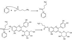
Figure 4. Reaction mechanism in the acylation of anthocyanins.
Table 4
Extract weights in UHT milk for samples preparation
Samples | Description | Weight (g) |
M1 M2 M3 M1R M2R M3R | UHT milk UHT milk with anthocyanin extract UHT milk with acylated anthocyanins Refrigerated UHT milk Refrigerated UHT milk with anthocyanin extract Refrigerated UHT milk with acylated anthocyanin extract | - 0.1029 ± 0.005 0.1035 ± 0.003 - 0.1035 ± 0.006 0.1030 ± 0.005 |
Then, the samples were prepared in 50 mL tubes and covered with aluminum foil to prevent photodegradation. After an 18-day storage period at room temperature, all three samples exhibited high stability, showing minimal fluctuations in both color intensity and pH values. The samples at room temperature formed clots, and the samples M1 and M2 developed a yellowish coloration. The sample M3 remained white with a smaller number of clots formed on the surface 18 days after the extracts were added. Meanwhile, the samples under refrige-ration at around 4 ºC maintained their color and did not form clots until 28 days after their preparation. After 80 days, precipitate was observed in sample M1R and in a lesser amount in sample M2R, while sample M3R did not form precipitate.
Tests were carried out at room temperature for the second time, where after 7 days the formation of two phases and a rancid odor were observed in M1 and M2, while in M1 a yellowish layer formed on the surface. M3 showed no alteration and had the characteristic smell of milk. On day 19, greater rancidity was observed in the milk samples, more so in samples M1 and M2. In sample M3, a thin yellow layer formed, and the rest of the milk content remained uniform and white.
An average density of 1.037 g/mL and pH of 6.3 were obtained, close to reported by Daszkiewicz et al. (2024) of 1.033 g/mL and 6.6, respectively. The acylated anthocyanins were found to be not completely soluble in UHT milk. The color change and formation of clots in the milk are due to its deterioration and decrease in pH, among other factors. The qualitative tests carried out over time demonstrated that the UHT milk sample with acylated anthocyanins (M3) showed greater stability compared to those containing non-acylated anthocyanin extract M2 and M1. In these, better preservation of sensory properties was observed, evidenced by a fresher color and smell in most of its content, as well as lower levels of coagulation and rancidity.
3.9 Fatty acid profile by gas chromatography FID
The fatty acid profile analysis was carried out using gas chromatography coupled to a flame ionization detector GC-FID with a 30 m column, 0.25 I.D (mm), 0.2 µm thickness. Table 5 shows the weights in grams and the fat content of each sample.
The determination of the fatty acid profile was carried out by gas chromatography with flame ionization detection (GC-FID), following the AOAC 996.06 method (2023). For the analysis, three milk samples stored at room temperature were prepa-red: M1, composed of 200 mL of UHT milk without additives; M2, consisting of 200 mL of UHT milk and 0.412 g of anthocyanins; and M3 with 200 mL of UHT milk and 0.412 g of acylated antho-cyanins.
Table 5
Total fats of UHT milk samples M0, M1, M2, M3
Samples | % Fat |
M0 M1 M2 M3 | 3.13 ± 0.02 2.84 ± 0.06 2.72 ± 0.04 1.14 ± 0.07 |
M0: Blank, UHT milk day zero; M1: UHT milk; M2: UHT milk with anthocyanin extract; M3: UHT milk with acylated anthocyanin solid extract.
A fresh UHT milk sample (M0) was used as a reference. The comparison of the fatty acids detected as a percentage of interest is shown in Table 6. The chromatograms corresponding to samples M0 and M3 are presented in Figures 5 and Figure 6, respectively. Samples M1, M2, and M3 were stored for 7 days in amber jars at room temperature before being analyzed.
Regarding the lipid composition, sample M1 (UHT milk without anthocyanins) had 2.84% total fat, of which 71.35% corresponded to saturated fatty acids. Sample M2 (milk with anthocyanins) showed a content of 2.72% total fat, with 72.15% saturated fatty acids. Finally, sample M3 (milk with acylated anthocyanins) contained 1.14% total fat, of which 72.19% were saturated.
Table 6
Comparison of the percentage % of fatty acids by samples
Fat acid | M0 | M1 | M2 | M3 |
Caproic Caprylic Lauric Palmitoleic Palmitic Oleic Stearic | 1.01±0.05 0.83±0.01 3.04±0.04 1.68±0.03 34.22±0.06 23.48±0.05 13.22±0.02 | 2.36±0.03 1.93±0.04 3.68±0.02 1.62±0.02 33.39±0.08 19.046±0.01 10.38±0.04 | 2.13±0.06 1.52±0.05 3.57±0.07 1.51±0.01 33.99±0.05 19.36±0.03 10.61±0.03 | 1.76±0.03 1.40±0.04 3.55±0.05 1.76±0.03 34.62±0.07 18.05±0.04 11.34±0.02 |
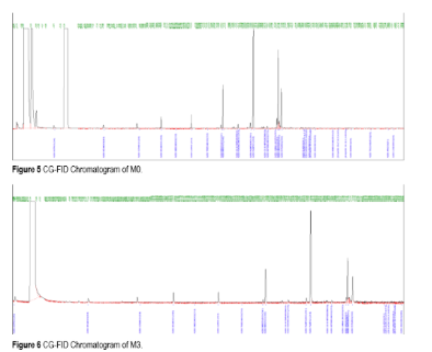
It was observed in Table 5 that, although sample M3 showed a lower total fat content, it managed to preserve saturated fats in a higher proportion compared to the others, as seen in Table 6. In addition, it showed superior stability in lauric and stearic fatty acids, with the lowest variations (16.8% and 14.2%, respectively). Likewise, caproic and caprylic acids showed increases of 74.0% and 68.8%, suggesting greater antioxidant protection against lipid oxidation. Palmitic and palmitoleic acids also showed minimal variations of 1.2% and 4.8%, respectively. Compared to samples M2 and M3, a similar percentage % area was observed with palmitic and oleic acids (Table 6). The propagation of free radicals of fatty acids should be stopped by incorporating acylated anthocyanin as an antioxidant since it exhibits a behavior as an effective electron donor. The acylated anthocyanin reacts with the free radical generating a phenolic radical, in addition to forming a hydroperoxide, without directly affecting the structure of the original fatty acid (Figure 7). This inhibitory effect suggests that acylated anthocyanins are capable of both quenching initial oxidative chain reactions and preventing the accumulation of secondary oxidation metabolites.
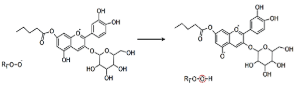
Figure 7 Reaction of the inhibition of the oxidation of a fatty acid by the acylated anthocyanin.
4. Conclusions
It was established that the incorporation of acylated anthocyanins from blueberry (Vaccinium corymbosum) improves the stability of UHT cow's milk, surpassing the effectiveness of non-acylated extracts in preserving organoleptic properties. Analyses by GC-FID confirmed that the acylated extract inhibits lipid oxidation by maintaining a higher proportion of the saturated fat fraction, with minimal variation in the concentrations of lauric, stearic, caprylic, and caproic acids, which validates the antioxidant potential of these modified molecules. These results benefit the dairy and agro-industrial industry by offering a natural and technologically superior alternative to extend the shelf life of products sensitive to oxidative degradation, ensuring greater nutritional value for the consumer.
Acknowledgment
Sincere gratitude to the Universidad Nacional Mayor de San Marcos for providing access to the laboratory facilities, Dr. Marcos Loroño for his support and expertise in computational chemistry, and the TYPSA laboratory for the use of the GC-FID equipment, which was crucial to the development of this article.
References
Association of Official Analytical Chemists. (2023). Official method 996.06: Fat (total, saturated, and unsaturated) in foods. En Official methods of analysis of AOAC International (17th ed.). AOAC International.
Codex Alimentarius. (2023). Norma general para los aditivos alimentarios (CODEX STAN 192-1995).
Daszkiewicz, T., Florek, M., Murawska, D., & Jabłońska, A. (2024). A comparison of quality of ultra-high-temperature milk and its plant-based analogs. Journal of Dairy Science, 107(12), 10299-10309. https://doi.org/10.3168/jds.2024-25098
Falcone, G., Mazzone, M., Cesa, S., Cairone, F., Carradori, S., Cellini, F., Chiavaroli, A., Orlando, G., Libero, M., Perrotti, V., Di Marcantonio, M., & Mincione, G. (2025). The anthocyanin-enriched extract of Vaccinium corymbosum exerted in vitro and ex vivo antiproliferative and anti-inflammatory activities. Journal of the Science of Food and Agriculture, 5(5), 199-211. https://doi.org/10.1002/jsf2.230
Godlewska, K., Pacyga, P., Najda, A., & Michalak, I. (2023). Investigation of chemical constituents and antioxidant activity of biologically active plant-derived natural products. Molecules, 28(14), 5572. https://doi.org/10.3390/molecules28145572
Johnson, J., El Orche, A., & Naiker, M. (2022). Prediction of antho-cyanin content and variety in plum extracts using ATR-FTIR spectroscopy and chemometrics. Vibrational Spectroscopy, 121, 103406. https://doi.org/10.1016/j.vibspec.2022.103406
Liu, Y., Tao, C., Liu, M., Pan, Y., & Lv, Z. (2018). Effect of temperature and pH on stability of anthocyanin obtained from blueberry. Journal of Food Measurement and Characterization, 12, 1744–1753. https://doi.org/10.1007/s11694-018-9789-1
Lock, O. (2016). Investigación fitoquímica: Métodos en el estudio de productos naturales (3.ª ed.). Pontificia Universidad Católica del Perú.
Taghavi, T., Patel, H., & Rafie, R. (2021). Comparing pH differential and methanol-based methods for anthocyanin assessments of strawberries. Food Science & Nutrition, 10(7), 2123-2131. https://doi.org/10.1002/fsn3.2065
Teng, H., Mi, Y., Deng, H., He, Y., Wang, S., Ai, C., Cao, H., Zheng, B., & Chen, L. (2022). Inhibitory effect of acylated anthocyanins on heterocyclic amines in grilled chicken breast patty and its mechanism. Current Research in Food Science, 5, 1732-1739. https://doi.org/10.1016/j.crfs.2022.09.011
Vyas, P., Kalidindi, S., Chibrikova, L., Igamberdiev, A. U., & Weber, J. T. (2013). Chemical analysis and effect of blueberry and lingonberry fruits and leaves against glutamate-mediated excitotoxicity. Journal of Agricultural and Food Chemistry, 61(32), 7769–7776. https://doi.org/10.1021/jf401158a
Wang, P., Liu, J., Zhuang, Y., & Fei, P. (2022). Acylating blueberry anthocyanins with fatty acids: Improvement of their lipid solubility and antioxidant activities. Food Chemistry X, 15, 100420. https://doi.org/10.1016/j.fochx.2022.100420
Yang, Z., Celik, M., & Arakawa, H. (2023). Challenges of Raman spectra to estimate carbonyl index of microplastics: A case study with environmental samples from sea surface. Marine Pollution Bulletin, 194, Part A. 115362. https://doi.org/10.1016/j.marpolbul.2023.115362